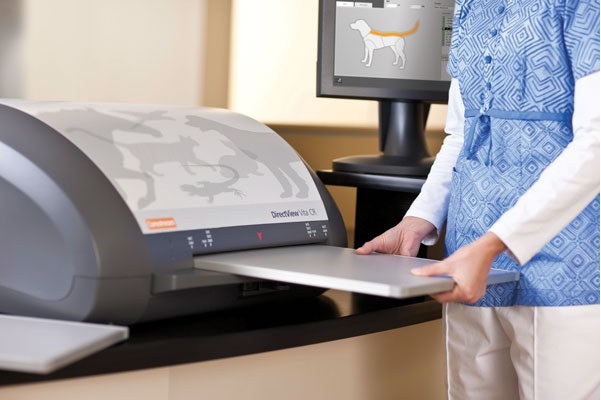
Non-Destructive Testing (NDT) is essential for hospitals to comply with industry regulations. Numerous imaging clinics and hospitals still rely on X-ray testing to diagnose critical illnesses in patients. If you want to update your hospital’s NDT processes, opting for Computed Radiography (CR) can be beneficial. CR has numerous benefits compared to conventional X-rays.
The CR Systems are rapidly replacing traditional film-based radiography in healthcare settings. But what exactly is CR, and how it works? Let’s explore what CR systems are and how they work.
What is Computed Radiography?
Computed Radiography is a type of digital radiography that replaces conventional X-ray imaging. Unlike traditional X-ray devices that use film, CR systems rely on phosphor imaging plates for capturing the images. After exposing the plate to radiation, it captures the test object’s image. Moreover, in CR, there is no need to process the image in a darkroom. The CR System converts the image into a digitalised version for analysis. The technician can make adjustments to analyse the image properly.
The Working Mechanism of the CR System
The working mechanism of the CR System involves the following stages.
● Exposure
Just like in traditional X-rays, the technician positions the patient to direct radiation through their body. As the X-ray beams pass through the body, they create a pattern of internal structures.
● Capturing the Image
In CR, a phosphor imaging plate is used. The technician exposes this plate to X-rays. The phosphor layer captures and stores the X-ray energy. This helps create a latent image or a pattern of electrons in the phosphor layer’s crystal structure.
● Analysis of the Plate
The reader in the CR System plays a crucial role in analysing the plate. The reader uses a laser beam to scan the plate. Nowadays, most manufacturers of CR Systems use laser beams to ensure rapid plate analysis.
● Converting the Image
The next stage is converting the image to digital form. The photomultiplier tube in the CR reader detects the laser beams and converts them into electrical signals. The analogue signal is then digitised, creating a raw digital image that represents the original X-ray pattern.
● Processing the Image
The computer system in the CR device processes the digital image to make it interpretable to doctors and technicians. Moreover, the technician has the flexibility to enhance the contrast, sharpen the edges, reduce noise from the picture, etc. These are critical enhancements that help improve image quality. Moreover, they assist the radiologists in visualising an array of anatomical structures.
Storage
A high-resolution monitor displays the processed image for the radiologist to analyse. The PACS stores the digital image for easy retrieval and sharing across the healthcare facility’s ecosystem. Undoubtedly, Computed Radiography is playing a crucial role in digitising medical imaging. The CR Systems offer a more flexible, efficient, and environmentally friendly approach to diagnostic imaging in healthcare facilities. Understanding the working mechanism of the CR Systems is vital when transitioning from film. Armed with this knowledge, you can make informed decisions and provide better care to patients.


